In traditional pathology, the pathologist, glass slide specimen, and the microscope all have to be in the same room together.
With cancer cases increasing and the pathology workforce declining, this method is untenable. In order to reach geographically distant pathologists and specialists, glass slides have to be physically mailed to the pathologist, leading to delay, breakage, and loss.
This method is costly from several perspectives. Pathologists must incur overhead and lost productivity due to travel, mailing glass slides is expensive and risky, and the delay in returning results, especially to a rural or remote community, can affect a patient's diagnosis and treatment.
We provide solutions that address these challenges. Our telemicroscopy systems improve patient care, increase efficiencies for pathologists, and reduce the overall cost of healthcare.
We are proud to innovate, design, and manufacture Mikroscan systems from our office headquarters in Carlsbad, CA. Our quality management system also meets the International Organization for Standardization (ISO) regulatory requirements for medical devices. We are ISO 13485:2016 certified for the design, development, sale, service, and distribution of whole-slide scanning and remote-controlled microscopy devices and applications for pathology.
Co-location and the reliance on glass slides are also barriers to researchers working on new discoveries. To address this problem, we provide digital pathology systems that create whole-slide digital images of pathology slides. This technology aids researchers who can perform quantitative analysis on specimens using image analysis algorithms specifically created for their application and collaborate with many multi-disciplinary teams, allowing them to make more advanced discoveries in less time. Educators can also use digital slide images to teach remote and online students using an archive of rare or interesting cases.
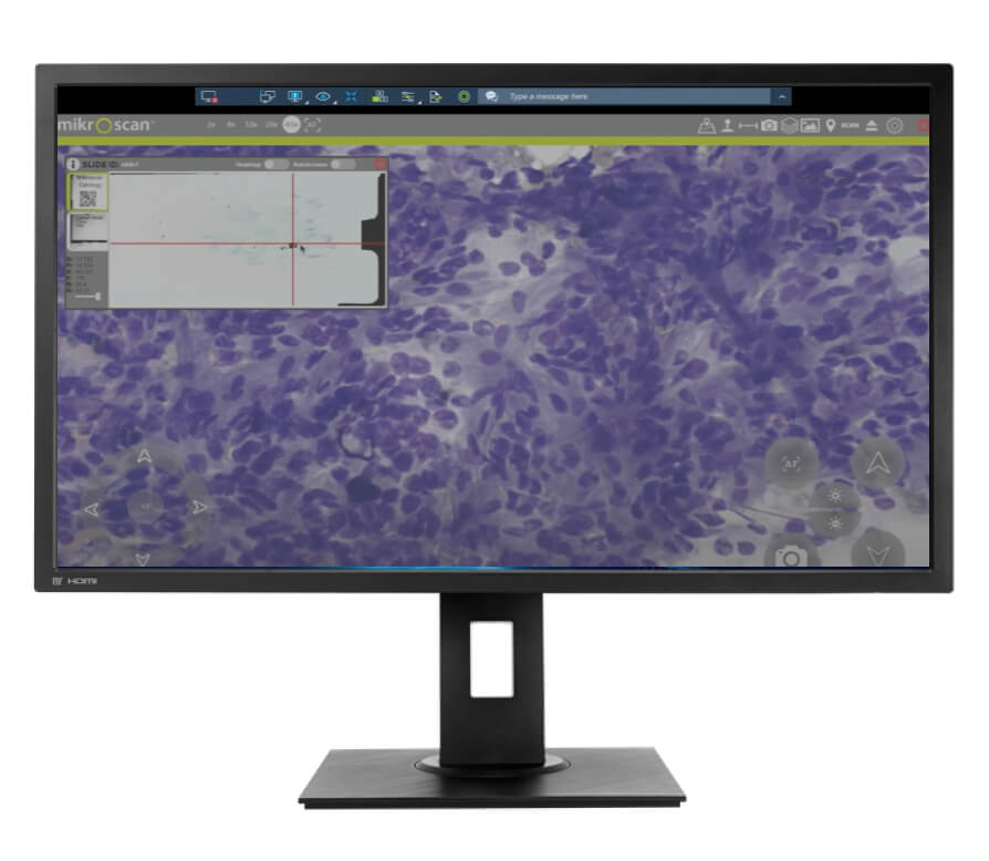
Our live telemicroscopy technology removes the requirement for co-location, allowing pathologists to log in remotely and provide the pathology support needed to diagnose and treat patients. By removing the requirement for proximity, patients can access the best possible subspecialty expertise from anywhere in the world.
Mikroscan enables patients to get the right diagnosis from the right expert at the right time.
Our technology improves the quality of patient care by connecting pathologists and specialists to remote and rural communities who often don't have many, if any, pathology services near them. Without our technology, these patients wouldn't receive the same level of care as those who live in a suburban or urban setting, close to hospitals and academic medical centers.
When pathologists use our Class I robotic microscope to remotely read slides, patients receive faster, more accurate results, and don't have to be burdened by the long wait caused by mailing glass slides to a pathologist.
They are spared having to undergo repeat biopsies and surgeries when it is discovered that there wasn't enough diagnostic material in their initial samples. Advances in remote on-site evaluation of adequacy (ROSE) enable cytopathologists to confirm the presence of diagnostic material in biopsy specimens while the patient is still in the office. The ability to provide immediate frozen section support while the patient is still in the operating room also greatly reduces the frequency of return visits and repeat surgeries.
Not only is the process more expedient and less stressful for the patient, but timely and more accurate results lead to a better outcome.
The pathology workforce is declining as cancer cases are rising. Needing to be physically present for every review places an unnecessary burden on these critical healthcare professionals.
The ability to review slides and perform primary diagnosis using a Class I telemicroscopy device, from anywhere, creates exponential efficiencies for busy pathologists.
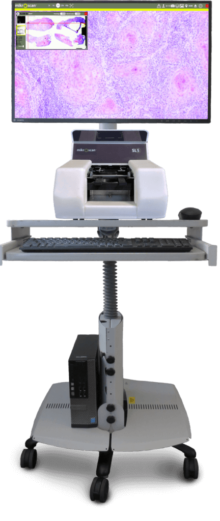
Our live telemicroscopy system operates just like a microscope, so it fits in seamlessly with the pathology workflow, and pathologists feel immediately comfortable using it. Implementation also doesn't require extensive infrastructure changes, so setup is easy and cost-effective, allowing pathologists to hit the ground running.
This increased efficiency also allows pathologists to expand their reach and generate more revenue.

Providing remote pathology services also greatly reduces the cost of healthcare.
In traditional pathology, pathologists have to travel to each site—some of them hours away—which is time-consuming and costly. Mailing slides back and forth is also expensive and causes unnecessary delay.
When hospitals employ live telemicroscopy, they experience a reduction in re-operation rates, repeat biopsies, and diagnostic errors.
Using ROSE, repeat biopsy rates have been shown to decrease by 20%, a $1.5K savings per biopsy1. The routine use of frozen sections accelerates margin assessment and reduces re-operations in any number of cancer types, such as oral, gynecological, dermatological, and lymph nodes. In breast cancer, it has been shown to reduce re-operations by 25%, an $18.2M savings2.
Our technology decreases these costs and delays, allowing pathologists to return more accurate results more quickly and at less overall expense.

It is imperative that we seek out solutions that help address the shift in reimbursement methodologies toward improved outcomes, reduced costs, and decreased risks. Mikroscan’s technology is a great tool for pathology departments to accomplish these objectives.
Keith Kaplan, MDPathologist |
We have a family of products to meet your pathology needs, from telemicroscopy systems to low- and medium-throughput digital pathology systems.
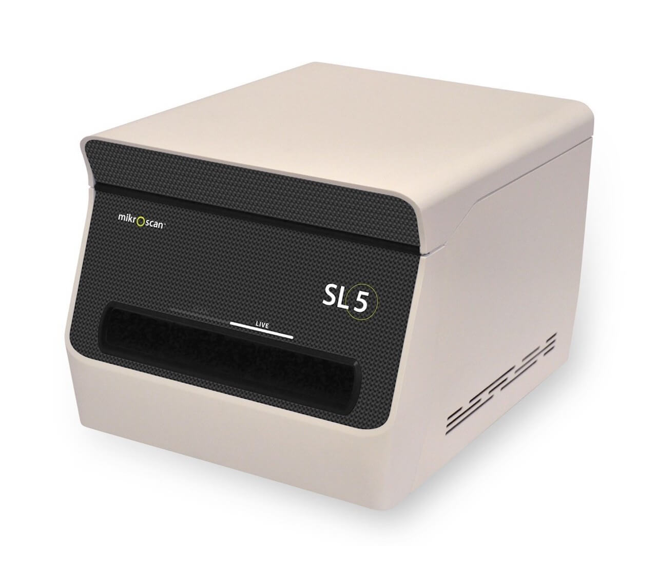
The SL5 Dual-Mode Real-Time Telemicroscopy and Digital Pathology System is the most compact, portable, and affordable telemicroscopy and digital pathology instrument on the market. It provides the added advantage of real-time telemicroscopy along with a completely separate mode for traditional "scan, store, and share" digital pathology applications. The SL5 contains a complete set of objectives—2X, 4X, 10X, 20X, and 40X—for true magnification of a wide range of applications and uses.

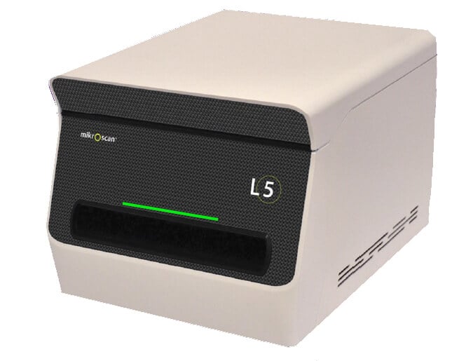
With the L5, you can be anywhere in the world and review a glass slide as though you were in the same room.
Using 5 high-quality microscope objectives, you will have complete control over slide navigation, focus, and illumination just like with a traditional brightfield microscope. You choose the planes of focus and areas of interest, not the system. Unlike with other systems, you can also control illumination from where you are to boost light for better examination of a thick specimen.

Trends in diagnostic interpretations reveal areas of improvement for current quality practices and provide vital opportunities for continuous improvement, patient safety, and positive outcomes. Read our case study to find out how to improve diagnostic proficiency in anatomic pathology.